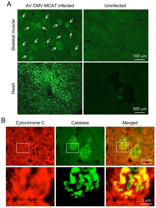
Figure 2. Systemic AV.RSV.MCAT infection leads to mosaic catalase expression in the mitochondria in striated muscles.
A, Representative catalase immunofluorescence staining photomicrographs of AV.RSV.MCAT infected and uninfected skeletal muscle (top panels) and heart (bottom panels). Arrow, AAV transduced skeletal muscle myofiber. B, Representative double immunofluorescence staining photomicrographs of AV.RSV.MCAT infected heart. Bottom panels are high magnification images of the boxed region in respective top panels. Cytochrome C marks mitochondria (red color). Catalase is in green color. Yellow color in merged images reveals mitochondrial catalase expression.

