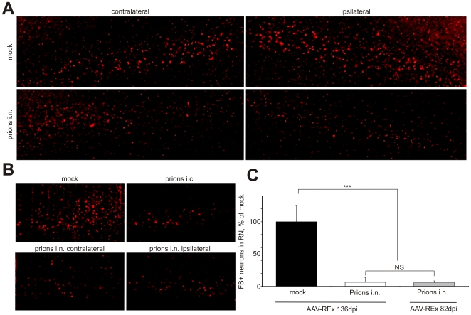
Figure 5. Reduction of DsRed-Express-positive neurons in motor cortex.
(A and B) Bilateral reduction of REx-positive neurons in the motor cortex (MC) of wild type (wt) mice upon prion challenge into the right sciatic nerve (prions i.n.) or intracerebrally (prions i.c.), as compared to mock controls, shown with confocal microscopy (A) and ultramicroscopy (B). (C) Quantification of REx-positive neurons on confocal images of MC shows a 94±3% (n = 4) reduction of tracer-positive cells in the MC of wt mice inoculated with prions i.n. at the disease onset (AAV-REx application at 136 days post prion inoculation, dpi) and a 95±1% reduction (n = 3) at 50% of incubation time (AAV-REx application at 82 dpi). Unpaired t-test was done using GraphPad Prism software. *** – P<0.0001. NS – non-significant. Scale bars: 100 µm.

