Figure 2.
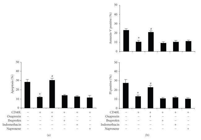
Oxaprozin-induced apoptosis is independent on COX inhibition. Monocytes were incubated for 48 hours with control medium or 200 ng/mL CD40L plus 1 μg/mL CD40L enhancer in the presence or absence of 100 μM oxaprozin, 100 μM ibuprofen, 100 μM indomethacin, or 100 μM naproxene. (a) Assessment of apoptosis by acridine-orange-stained slides under fluorescence microscopy. Data are express, as percentages of apoptotic cells on total number of cells counted (mean ± SEM, n = 4; apoptosis in absence versus in presence of CD40L: *P < .05; apoptosis in presence of CD40L versus CD40L plus oxaprozin: #P < .05; apoptosis in presence of CD40L versus CD40L plus other NSAIDs: N.S.). (b) Assessment of apoptosis by using Annexin V and Propidium Iodide (PI) binding assay (mean ± SEM, n = 3; apoptosis in absence versus in presence of CD40L: *P < .05; apoptosis in presence of CD40L versus CD40L plus oxaprozin: #P < .05; apoptosis in presence of CD40L versus CD40L plus other NSAIDs: N.S.).

