Figure 1.
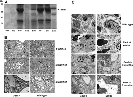
Glomerular disease is present in Par4−/− mice. (A) A total of 4 μl of urine from 3-mo-old WT (WT1 and WT2) and Par4−/− (KO1 through 6) mice was suspended in Laemmli buffer and subjected to SDS-PAGE. Coomassie staining detected proteinuria in Par4−/− mice but not their WT counterparts. A band corresponding to albumin is shown (68 kD; black arrow). The variability in proteinuria resolved with increasing age of mice. (B) Hematoxylin and eosin staining of Par4−/− and WT glomeruli at 2 wk (a and b), 3 mo (c and d), and 6 mo (e and f). At 2 wk, only mild mesangial hypercellularity and glomerular enlargement is present in Par4−/− glomeruli, with preservation of the tubulointerstitial compartment (a). At 3 mo, marked mesangial expansion, glomerulosclerosis, and widening of the subcapsular space is evident, with some capsular adhesions (c). Significant disease progression occurs by 6 mo, with widespread scarring and atrophy of glomeruli, tubular atrophy, and pseudocysts containing proteinaceous material (arrow; E). In 25% of mice, coexisting renal tubular cysts were lined by simple epithelium (*), which also progressed in severity with age. Glomeruli and renal tubules were normal at all ages in WT controls (B, D, and F). (C) Par4−/− glomeruli were also abnormal at the ultrastructural level (c through h). In 2-wk-old mice (c and d), there is discontinuous podocyte foot process effacement and mild cell body swelling, but the GBM is normal. By 3 mo, there is marked effacement and swelling with hyaline deposits and vacuoles, glomerulosclerosis, mesangial expansion, and patchy blebbing of the GBM (e and f). By 6 mo, podocyte swelling, sclerosis, and foot process fusion is extensive, with diffuse thickening, corrugation, and denudation of the GBM. The mesangial matrix is folded and collapsed (g and h). By contrast, normal podocyte and glomerular architecture is seen in WT mice and littermates (a and b). Podocyte foot processes are well demarcated, and the mesangium is normal with preservation of the urinary space and no tubular cysts. Magnifications: ×40 in B; ×300 in C (a, c, e, and g); ×6000 in C (b, d, f, and h).

