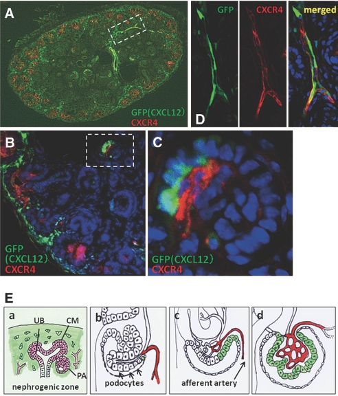
Figure 3.
Spatial relationship between CXCL12 and CXCR4 in developing kidney. Kidney sections from CXCL12/GFP knock-in mice were stained red (Alexa555) for CXCR4. (A and B) Appearance of whole kidney section (A) and magnified image (B) demonstrates that CXCL12-positive stromal cells surround CXCR4-positive developing nephrons. (C) Higher magnification of the glomerulus in the dotted lines in B suggests that some podocytes express CXCL12 and that glomerular endothelial cells just adjacent to the podocytes express CXCR4. (D) Higher magnification of the region in the dotted lines in A of GFP (left) and CXCR4 staining (middle) in the interlobular arteries. Merged image (right) indicates that at least some interlobular arteries express both CXCL12 and CXCR4. (E) Summary of the expression of CXCL12 (green) and CXCR4 (red) in the developing early nephron (a) and glomeruli (b through d). See the Discussion section for more details. Magnifications: ×100 in A; ×400 in B; ×600 in C and D.

