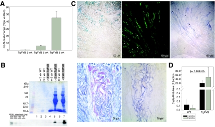
Figure 3.
Induction of uNGAL. (A) NGAL real-time PCR in TgFVB kidneys from 3-, 6-, and 8-wk-old mice. Data were normalized for NGAL expression in age- and sex-matched WT FVB/N littermate mice. (B) Coomasie blue stained gels and immunoblots of uNGAL in 3-, 6-, and 8-wk-old TgFVB and WT littermate mice. uNGAL increased between 6 and 8 wk in TgFVB mice, whereas proteinuria remained constant. (C) Sections of 8-wk-old TgFVB mouse kidneys. From left to right: Prussian blue staining demonstrates iron accumulation in cortical proximal tubules. Aquaporin-2 immunocytochemistry marks medullary collecting ducts while in situ hybridization in an adjacent section reveals NGAL expression (blue) in dilated collecting ducts (asterisk represents dilated tubules). Higher power demonstrates cast formation (periodic acid—Schiff stain, purple) and NGAL expression (blue) in two adjacent sections. (D) Increasing number of cysts per unit area in TgFVB kidneys with aging. TgFVB differed significantly from WT kidneys (n = 14, P = 0.00074 at 4 wk; n = 14, P = 0.001 at 8 wk).

