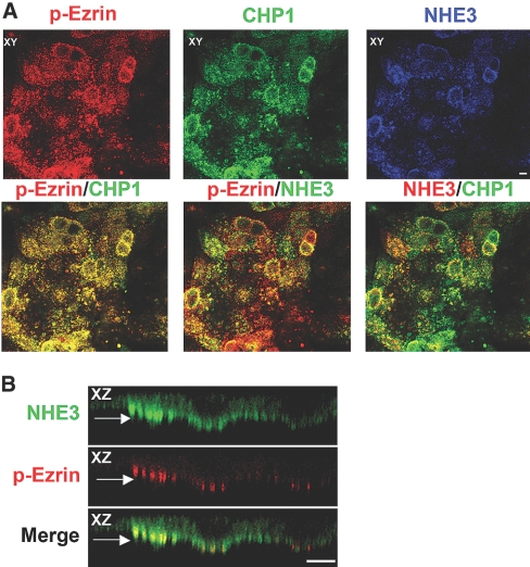
Figure 3.
Colocalization of NHE3 and CHP1 with endogenous phosphorylated ezrin. Localization of endogenous NHE3, CHP1, and phosphorylated ezrin by confocal microscopy. NHE3 was stained with #3H3 antiserum, CHP1 with anti-CHP polyclonal antibody, and phosphorylated ezrin with T567 phospho-specific polyclonal antibody. Colocalization of NHE3, CHP1, and phosphorylated ezrin is shown. Colocalization of two of the three molecules is indicated by the merged yellow color. Pseudocolor of green or red was assigned to NHE3 as indicated in the figure. XZ cross-sections and XY face views are highlighted. (A) Colocalization of endogenous NHE3, CHP1, and phosphorylated ezrin (p-Ezrin). (B) Detail of endogenous NHE3 and p-Ezrin colocalization in apical clusters as indicated by the arrow in the XZ plane. Scale bar, 2 μm.

