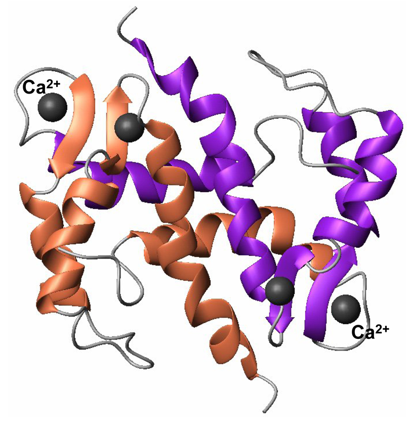
Fig. 1.
MolMol representation of the three-dimensional structure of S100A13 (PDB ID 1CXJ) bound to calcium. S100A13 is a dimer and each monomeric unit consists of four helices arranged in to two EF-hand motifs. Each EF hand motif binds to a Ca2+ ion. Residues involved in the calcium binding site form a beta-sheet type of structure.

