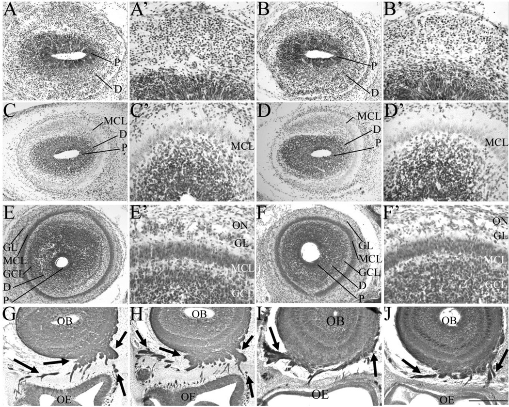
Fig. 2.
A–J: The glomerular layer is hypocellular in the absence of Sall3. Nissl staining of coronal sections of the olfactory bulb at E14.5 (A–B′), E16.5 (C–D′), and P0.5 (E–F′) and Tuj1 staining at E16.5 (G,H) and P0.5 (I,J) in control (A,A′,C,C′,E,E′,G,I) and Sall3−/− (B,B′,D,D′,F,F′,H,J) animals. Control animals represent images of either Sall3+/+ or Sall3+/− animals. No alterations in early olfactory development were observed in Sall3 mutant animals (B,B′,D,D′). The GL appeared hypocellular at P0.5 in Sall3-deficient animals (F′). The MCL, GCL, and GL were identifiable under high power, by cellular characteristics described in Materials and Methods. Innervation of the olfactory bulb by the olfactory nerve (arrows, G–J) was not altered in Sall3 mutant animals (H,J). P, progenitor populations; D, differentiating field; GCL, granule cell layer; MCL, mitral cell layer; GL, glomerular layer; ON, olfactory nerve; OB, olfactory bulb; OE, olfactory epithelium. Scale bar = 100 µm for A′,B′,C′,D′,E′,F′; 200 µm for A,B; 300 µm for C,D,G,H; 350 µm for E,F,I,J.

