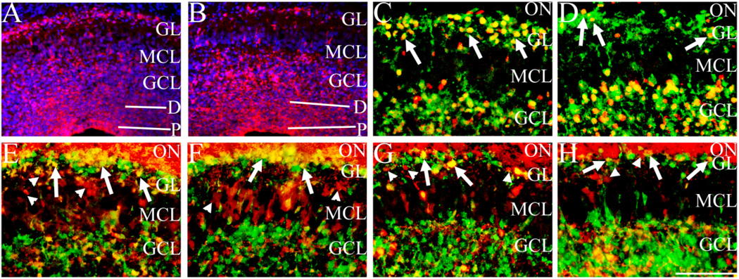
Fig. 8.
Sall3 is not required in a cell-autonomous manner for glomerular olfactory interneuron maturation. β-Galactosidase expression (red) in Sall3+/− and Sall3−/− animals at P0.5 (A,B). Expression is observed in progenitor cells, differentiating cells, GCL, MCL, and GL (A,B). Sections were counterstained with DAPI (blue). In Sall3 mutant animals, a decrease in the number of β-galactosidase-expressing cells in the GL and an increase in β-galactosidase-expressing cells in the GCL were observed (B). Pax6 (red; C,D), calretinin (red; E,F), and calbindin (red; G,H) coexpression with β-galactosidase (green) in the P0.5 olfactory bulb in Sall3+/− (C,E,G) and Sall3−/− (D,F,H) animals. Coexpression of β-galactosidase was observed with the majority of Pax6 (arrows, C) cells in the GL. However, a mix of calretinin+ β-galactosidase+ (arrows, E) and calretinin+ β-galactosidase− (arrowheads, E) and calbindin+ β-galactosidase+ (arrows, G) and calbindin+ β-galactosidase− (arrowheads, G) was observed in the GL of Sall3+/− animals. In Sall3 mutant animals, although the number of Pax6-, calretinin-, and calbindin-positive cells in the GL is decreased (D,F,H), cells coexpressing β-galactosidase and Pax6 (arrows, D), β-galactosidase and calretinin (arrows, F), and β-galactosidase and calbindin (arrows, H) were observed. Calretinin and calbindin are additionally expressed by olfactory nerve axons, visible as staining superficial to the GL in E–H. P, progenitor populations; D, differentiating field; GCL, granule cell layer; MCL, mitral cell layer; GL, glomerular layer; ON, olfactory nerve; β-gal, β-galactosidase; CR, calretinin; CB, calbindin. Scale bar = 150 µm for A,B; 100 µm for C–H.

