
Figure 5. TNF-α-mediated CBP upregulation is dependent on NF-κB.
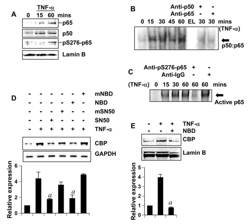
(A) Cerebellar granule neurons were treated with 10 ng/ml TNF-α for indicated times. Subsequently, nuclear extracts were isolated and 25μg nuclear extract was separated by gel electrophoresis and immunoblotted with anti-p65 and anti-p50 (both 1:1000 dilution). Membranes were stripped and reprobed with anti-phosphoS276-p65 (1:500) and anti-LaminB (1:1000) (loading control for nuclear extract). (B and C) Cerebellar granule neurons (B) and cortical neurons (C) were treated with 10 ng/ml TNF-α for indicated minutes. Then nuclear extracts (15μg) were used to perform EMSA with a labeled consensus κB probe. For supershift assay (rightmost lanes), 1μg anti-p65 and anti-p50 antibody (B) or 1μg anti-phosphoS276-p65 and normal IgG (C) was added to the binding mixtures. EL (B): empty lane. (D) Cerebellar granule neurons incubated with 2 μM SN50 (or mutated-SN50) or 5μM NBD peptide (or mutated-NBD peptide) for 2 hours were treated with 10 ng/ml TNFα for 18 hours followed by RT-PCR analysis. The relative expression of CBP (CBP/GAPDH) was measured after scanning the bands (lower panel). Results represent mean ± SD of three separate experiments. ap < 0.001 versus TNF-α. (E) Cerebellar granule neurons incubated with 5μM NBD peptide for 2 hours were treated with 10 ng/ml TNF-α for 24 hours. Nuclear extracts (25μg) were separated by gel electrophoresis and western blotted. The membranes were probed with anti-CBP antibody and then re-probed with anti-LaminB antibody. The relative expression of CBP (CBP/LaminB) was measured after scanning the bands (lower panel). Results represent mean ± SD of three separate experiments. ap < 0.001 versus TNF-α.