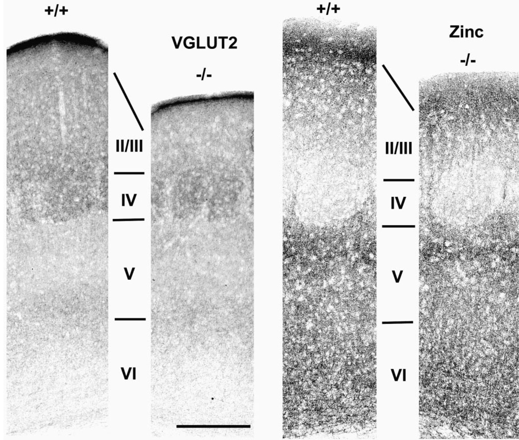
Figure 6.
Cortical circuits reflect cortical cytoarchitecture. Panels show pairs of coronal sections through barrel cortex of wild-type (+/+) and mutant (−/−) mice stained immunohistochemically for VGLUT2 (left pair of panels) or for synaptic zinc (right pair of panels). Each panel is centered on a layer IV barrel as determined from adjacent Nissl-stained sections. Note the complementary density and distribution of axon terminals stained by these two markers. Also note comparable staining patterns in wild-type and mutant cortex for layers IV through VI, and the attenuated thickness of layers II/III. Bar = 300 µm for all panels.

