Fig. 5.
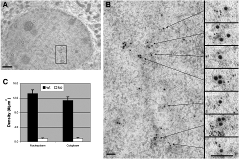
Double immunogold labeling and electron microscopy of L-FABP and PPARα binding sites in mouse liver. Antigenic sites of L-FABP were labeled with 6 nm gold particles and antigenic sites of PPARα were labeled with 15 nm gold. A: Multiple sites of colocalization in a control mouse hepatocyte were marked by circles in this low magnification image. This image focuses on the hepatic nucleus (darker circle which comprises most of the image) and some surrounding cytoplasm. B: The boxed region of the nucleus in (A) was enlarged and seven individual sites of colocalization were subsequently enlarged 2.5× to better visualize the localization of the 6 nm gold particles of L-FABP in proximity with the 15 nm gold particles of PPARα. C: Bar graph of the antibody-L-FABP labeling particle density in nucleoplasmic and cytoplasmic regions of hepatocytes obtained from L-FABP+/+ and L-FABP−/− mice. Bars = 1.0 μm in (A); 100 nm in (B). The images (A) and (B) were modified in Adobe Photoshop to adjust the brightness and contrast ( Curves ), remove random noise (“Despeckle”), and clarify positions of gold particles (“Unsharp Mask”).

