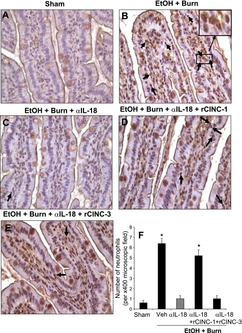
Fig. 5.
Intestinal neutrophil infiltration as determined by immunoperoxidase staining using anti-neutrophil antibody on day 1 after injury. Representative photomicrographs of intestine from all 5 experimental groups are shown (A–E). The sections were examined with Nikon Eclipse 50i microscope with a magnification of ×400. The sections were photographed with a camera attached to the microscope, and the images were digitally transferred into Photoshop. Inset in B shows digitally enhanced neutrophil infiltrated area. Along with immunopositivity, the presence of neutrophils was also confirmed by their distinct morphological nature with a multilobed nucleus. Each section was scanned for 3–4 microscopic fields, and the data thus obtained from more than 4 animals in each group were pooled and presented in F as means ± SE. *P < 0.05 compared with sham and EtOH + Burn + αIL-18. α, anti; Veh, vehicle.

