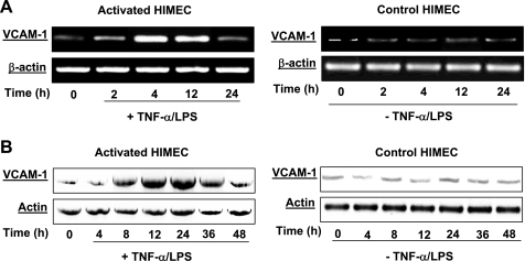
Fig. 1.
Effect of tumor necrosis factor (TNF)-α/lipopolysaccharide (LPS) on vascular cell adhesion molecule (VCAM)-1 mRNA and protein expression in human intestinal microvascular endothelial cells (HIMEC). The effect of TNF-α/LPS on VCAM-1 mRNA and protein expression was determined. A: detection of VCAM-1 mRNA in HIMEC by semiquantitative reverse transcriptase-PCR using specific primers. HIMEC does not constitutively express mRNA for VCAM-1. Stimulation of HIMEC with a combination of TNF-α (100 IU/ml) and LPS (1 μg/ml) led to marked upregulation of VCAM-1. VCAM-1 mRNA expression in HIMEC following TNF-α/LPS activation was time dependent; the maximum response was achieved by 4 h, which was lasted for 12 h and then returned to the basal level by 24 h (left). β-Actin served as an internal loading control. B: TNF-α/LPS enhanced VCAM-1 protein expression in HIMEC. VCAM-1 protein expression in HIMEC following TNF-α/LPS activation was assessed using Western blot analysis. Total cell lysates from cultured HIMEC stimulated with TNF-α (100 IU/ml) and LPS (1 μ g/ml) were subjected to SDS-PAGE and immunoblotted with VCAM-1 antibody. TNF-α/LPS activation of HIMEC significantly increased VCAM-1 protein expression by 12 h, was sustained through 24 h, decreased at 36 h, and returned to baseline by 48 h (left). Time-matched controls for VCAM-1 mRNA and protein are shown on right. The same blot was stripped and reprobed with an anti-actin antibody to ensure equivalent protein loading. Data shown are from one of three independent experiments.

