Fig. 3.
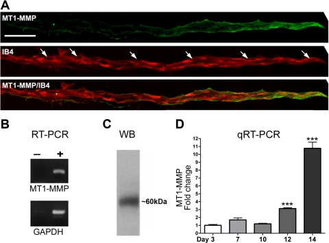
Membrane type 1 (MT1)-MMP expression in angiogenic outgrowths. A: confocal images of a representative neovessel from a collagen gel culture of rat aorta double-stained with an anti-MT1-MMP antibody (green) and an endothelial cell marker (IB4; red). Note: there is a gradient of MT1-MMP that is strongly expressed in the endothelial tip cells and barely detectable at the root of the neovessel. Arrows in the IB4-stained panel indicate nuclei (scale bar, 30 μm). B: photographs of ethidium bromide-stained gels showing PCR products from RT-PCR performed with (+) and without (−) reverse transcriptase on RNA isolated from rat aortic ring cultures and primers for MT1-MMP and GAPDH. C: photograph of a Western blot (WB) showing MT1-MMP expression in rat aortic cultures. A single band of ∼60 kDa was detected with anti-MT1-MMP antibody. D: quantitative real-time RT-PCR (qRT-PCR) of aortic ring cultures demonstrated progressive increase of MT1-MMP expression over time. Values are means ± SE; n = 3, ***P < 0.001.

