Fig. 6.
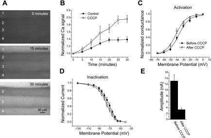
Effect of elevation of intracellular Ca2+ on sodium current in the absence of injury. A: images of muscle fibers (1–4) loaded with Fluo4 NW at the time of application of CCCP (0 min) and 15 and 30 min later. The increase in Fluo4 NW signal is the most rapid and largest in fiber 1. However, all 4 fibers have significant elevation of intracellular Ca2+. B: plot of the time course of increase in the normalized Ca2+ signal in control fibers following impalement versus fibers treated with CCCP but not impaled (n = 28). The increase in Ca2+ level was much greater following application of CCCP but had a similar time course to the increase following impalement. C: voltage dependence of activation before (solid squares) and after (open squares) treatment with CCCP. A shift in the midpoint of activation from −35.4 ± 1.3 mV to −39.7 ± 1.5 mV was present following application of CCCP (P < 0.05, n = 16). D: steady-state voltage dependence of inactivation of fibers before and after application of CCCP. The mean midpoint of inactivation shifted from −74.1 ± 2.0 mV to −78.1 ± 1.3 mV following application of CCCP (P < 0.05, n = 16). E: sodium current amplitude before and after application of CCCP. The same patch pipette was used to record current before and after CCCP for each fiber. The mean current amplitude fell by close to 75% (P < 0.01).

