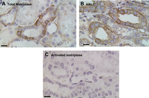
Fig. 3.
Distribution of matriptase and HAI-1 in human kidney. Paraffin-embedded human kidney specimens were stained by immunohistochemistry using mAbs against total matriptase (A), activated matriptase (C), or HAI-1 (B). Positive staining was observed as brown precipitates (diaminobenzidine), and nuclei were counterstained with hematoxylin. Bar = 20 μm.

