Fig. 4.
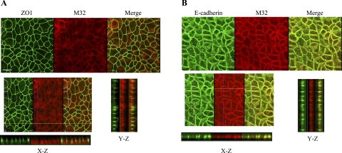
Subcellular localization of matriptase in polarized Caco-2 human intestinal epithelial cells. Caco-2 cells were grown and allowed to undergo differentiation on coverslips for 12 days. The polarized cells were fixed, permeabilized, and stained for matriptase with Alexa Fluor 594-conjugated mAb M32. The tight junction marker ZO-1 (A) and the adherens junctions marker E-cadherin (B) were costained with matriptase using FITC-conjugated antibodies to each protein. The staining was observed using a Nikon confocal microscope, and a series of images at different planes in the Z-axis were acquired. Representative images for matriptase, ZO-1, and E-cadherin from both staining studies are shown in the top panels, as indicated. The images in the X-Z- and Y-Z-axes were assembled from serial Z-section images and are shown in the bottom panels, as indicated. The scale bar represents 10 μm.

