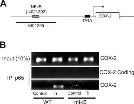
Fig. 5.
Ti particles stimulate NF-κB binding to the native COX-2 promoter. A: schematic of the COX-2 promoter showing the location of the NF-κB binding site and the expected PCR product in the chromatin immunoprecipitation assay. B: wild-type and mIκB FLS in subconfluent cultures were serum starved overnight and then treated with control medium or medium containing titanium (5 × 10⁶ particles/ml) for 4 h. The protein-DNA complexes were cross-linked, and genomic DNA was extracted by immunoprecipitation (IP) with p65 antibody. PCR was performed using primers flanking the consensus NF-κB-binding site on the COX-2 gene as shown in A. The input control consisted of PCR amplification of the COX-2 promoter obtained from total genomic DNA before immunoprecipitation. The coding control used primers located in the coding region of the COX-2 promoter (+5555 to +5654).

