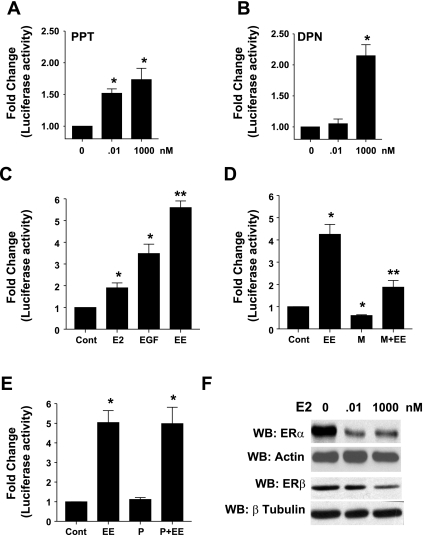
Fig. 2.
EGF selectively enhances ERα-stimulated prolactin (PRL) gene expression. GH3 cells transiently cotransfected with PRL reporter gene and control reporter gene (Cont) were treated with the indicated concentrations of PPT (A), DPN (B), or E2 (0.01 nM), EGF (5 ng/ml), or a combination (EE) (C) for 24 h, and normalized luciferase activity was determined as described in materials and methods. Data were calculated as fold change over control (arbitrary value of 1). Each value is the mean ± SE of 3 separate experiments, each performed in triplicate. *Significant difference from control. **Significant differences from E2 and EGF alone (P < 0.05). GH3 cells, transiently cotransfected with PRL reporter gene and control reporter gene were treated with vehicle or a combination of E2 and EGF (EE) either by themselves or in presence of the ERα-specific antagonist, MPP (100 nM) (D), or the ERβ-specific antagonist, PHTPP (100 nM) (E), for 24 h, and normalized luciferase activity was determined as described in materials and methods. Data were calculated as fold change over control (arbitrary value of 1). Each value is the mean ± SE of 3 separate experiments, each performed in triplicate. *Significant difference from control. **Significant differences from EE (P < 0.05). F: GH3 cells were treated with the indicated concentrations of E2 for 24 h. An equal amount of cell lysate was subjected to Western blotting (WB) with anti-ERα antibody (Ab) or anti-ERβ Ab to ensure equal loading blots were stripped and reprobed with either anti-actin Ab or anti-β tubulin Ab. Results shown are from a single experiment and are representative of 3 separate experiments.

