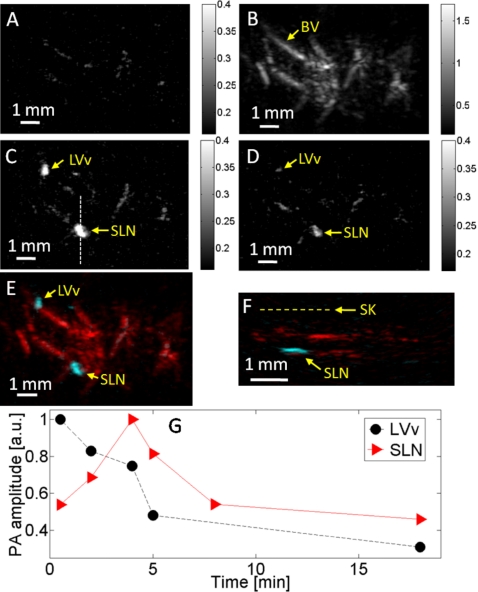
Figure 4.
Noninvasive in vivo photoacoustic mapping and dynamic monitoring of the sentinel lymph node in a rat. (a) Control photoacoustic MAP image acquired at 600 nm laser wavelength before Evans blue injection. (b) Control photoacoustic MAP image acquired at 584 nm, showing the subcutaneous vasculature. (c) Photoacoustic MAP image acquired at 600 nm 6 min after Evans blue injection. LVv, lymphatic valve; SLN, sentinel lymph node. (d) Photoacoustic MAP image acquired at 600 nm 15 min after Evans blue injection. (e) Composite photoacoustic MAP image. (f) Composite photoacoustic B-scan image corresponding to the dotted line in (c). SK, skin surface. (g) Evans blue dynamics in the rat lymphatic valve and SLN.

