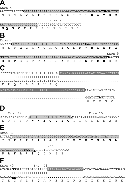
Fig. 2.
Additional splicing sites identified in cerebral artery CaV1.2 α1-subunits. Insertions are illustrated as black font shaded in light gray, with deletions as white font shaded with dark gray. *Stop codon. Amino acids introduced by insertions are indicated in bold font. Exon/exon junctions are presented as underlined bold font. A: 66-nt insert (155117087-155117024) between exons 4 and 5 in clone C4. B: 108-nt insert (155229797-155229690) between exons 4 and 5 in clone C8. C: 73-nt deletion (154984855-154984783) within exon 15 in clone C17. D: 21- nt insert (154984876-154984856) between exons 14 and 15 in clone C5. E: 71-nt insert (155174200-155174130) between exon 32 and 33 in clone B9. F: 18-nt deletion (154911308-154911291) within exon 41 in clone C16.

