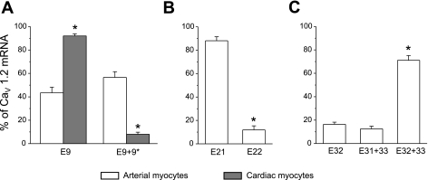
Fig. 3.
Real-time quantitative RT-PCR identifies relative expression of major CaV1.2 exon variants in isolated cerebral artery smooth muscle cells and cardiac myocytes. A: exon 9* expression is higher in cerebral artery smooth muscle cells (n = 6) than in cardiac myocytes (n = 5). *P < 0.05 when compared with exon 9 and 9* expression in arterial smooth muscle cells. B: in cerebral artery smooth muscle cells, CaV1.2 subunits preferentially express exon 21 (n = 4). *P < 0.05 when compared with exon 21 expression. C: exon 32+33 is the major splice variant in cerebral artery smooth muscle cells (n = 5). *P < 0.05 when compared with exon 32 and exon 31+33 expression. Each n is the calculated mean of real-time PCR experiments that were performed in triplicate.

