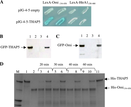
Fig. 1.
Thanatos-associated protein (THAP) 5 is an interactor and substrate of Omi/HtrA2 protease. A: interaction of Omi/HtrA2 with THAP5 protein in yeast. Yeast colonies were transformed with plasmids encoding the indicated baits and full-length THAP5 cloned into the prey vector. The specificity of THAP5 and Omi/HtrA2 interaction was verified using a closely related homolog, the HtrA1 protein. Blue color results are from a positive protein-protein interaction. B: interaction of Omi/HtrA2 and THAP5 in mammalian cells during apoptosis. Human embryonic kidney (HEK)-293 cells were plated in duplicates in 60-mm dishes and then transfected with either enhanced green fluorescent protein (EGFP), EGFP-THAP5, or green fluorescent protein (GFP)-Omi. Twelve hours after transfection, one plate was treated with 50 μM cisplatin to induce apoptosis, and one plate was used as control. Cell lysates were prepared as described in materials and methods. A polyclonal Omi/HtrA2 antibody was used to immunoprecipitate the endogenous Omi/HtrA2 and any associated proteins. The immunoprecipitated complex was resolved on SDS-PAGE and transferred to a polyvinylidene difluoride (PVDF) membrane, and the presence of GFP-THAP5 fusion protein was detected using a specific anti-GFP antibody. Lane 1 shows crude lysates of GFP-THAP5 transfected cells. Lane 2 shows coimmunoprecipitation (co-IP) lysates obtained from cells transfected with GFP empty vector. Lane 3 shows co-IP lysates obtained from cells transfected with GFP-THAP5 control cells. Lane 4 shows co-IP lysates obtained from cells transfected with GFP-THAP5 and then treated with cisplatin. C: the reverse experiment described in B. HEK-293 cells were now transfected with GFP-Omi and processed exactly as in B, except THAP5 antiserum was used in the co-IP and anti-GFP on the Western blot to detect the GFP-Omi. Lane 1 shows total cell lysates of GFP-Omi transfected cells. Lane 2 shows co-IP lysates obtained from cells transfected with GFP empty vector. Lane 3 shows co-IP lysates obtained from cells transfected with GFP-Omi control cells. Lane 4 shows co-IP lysates obtained from cells transfected with GFP-Omi and then treated with cisplatin. In both B and C, THAP5 was coprecipitated with Omi/HtrA2, but only in cells in which apoptosis was induced. D: THAP5 is cleaved by Omi/HtrA2 protease in vitro. Purified His-THAP5 was incubated with His-Omi134-458 at 37°C for the indicated time periods. For some assays, Omi/HtrA2 was preincubated with ucf-101 inhibitor 10 min before the addition of His-THAP5. The reactions were resolved on SDS-PAGE, and the gel stained with Coomassie blue. Lane 1: His-THAP5 control (400 ng); lane 2: His-Omi134-458 control (400 ng); lanes 3, 5, 7, and 9: His-Omi+His-THAP5 at different time points; lane 4, 6, 8, and 10: His-Omi + His-THAP5 + ucf-101 (50 μM); lane 11: His-THAP5 + ucf-101 control.

