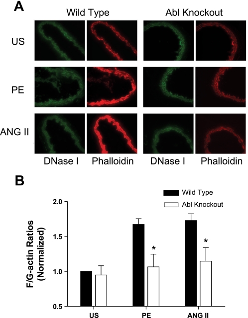
Fig. 3.
Enhancement of F-actin-to-G-actin (F/G-actin) ratios during PE or ANG II stimulation is inhibited in arteries from Abl−/− mice. A: representative images showing the effects of Abl deficiency on actin filament polymerization stimulated by agonists. Carotid arteries from Abl KO or WT mice were stimulated with PE (10 μM, 5 min) or ANG II (1 μM, 2.5 min), or they were unstimulated (US). Cryosections of these arteries were stained with Alexa 488-DNase I for G-actin (green) and with rhodamine-phalloidin for F-actin (red), and images were captured using a digital fluorescent microscope. B: F/G-actin ratios in response to agonist stimulation and the ratios of unstimulated tissues from KO mice are normalized to US values of arteries from WT mice. *P < 0.05, significantly lower F/G-actin ratios in Abl−/− arteries upon contractile stimulation compared with corresponding values obtained from Abl+/+ arteries. Values are means ± SE; n = 5 to 6.

