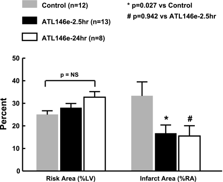
Fig. 3.
Infarct size as % of risk area (%RA). Myocardial risk area (left) and infarct size (right) in the experimental model of myocardial infarction. There is no difference in risk area between the 3 groups as measured by monastral blue dye. However, the control animals had a larger infarct size compared with ATL-146e-treated animals. There was no difference in infarct size between the animals that received a 2.5-h infusion and a 24-h infusion of ATL-146e. NS, not significant.

