Fig. 3.
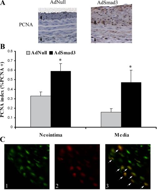
Smad3 overexpression increases cell proliferation in vivo. A: rat carotid arteries were infected with AdSmad3 (n = 5 rats) or AdNull (n = 5 rats) and, 14 days after injury, were stained for proliferating cell nuclear antigen (PCNA). Arrows indicate medial borders. B: PCNA index, as described in materials and methods, increased in both the neointima (AdNull, 0.33 ± 0.04; and AdSmad3, 0.59 ± 0.08) and the media (AdNull, 0.16 ± 0.04; and AdSmad3, 0.47 ± 0.13). *P < 0.05 (magnification, ×200). C: double immunofluorescent labeling for Smad3 and PCNA in neointima of injured rat carotid arteries: 1) Smad3-positive cells stained with fluorescein, 2) PCNA-positive cells stained with Texas red, and 3) superimposing the red and green images reveals that PCNA-positive cells are also Smad3 positive (white arrows) (magnification, ×400).

