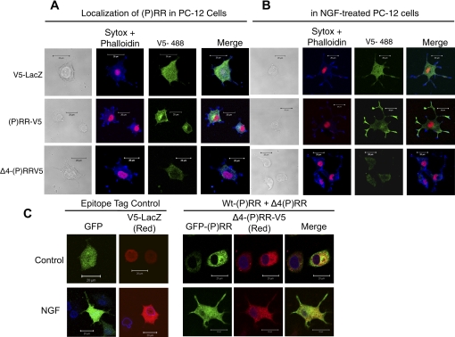
Fig. 5.
Cellular distribution of (P)RR and Δ4-(P)RR. Following transfection, the cells were treated without NGF (A) or with NGF (B) and subjected to immunofluorescence staining. V5-tagged proteins are stained with AlexaFluor-488 (green) and nuclei stained with Sytox-orange (red). Actin is stained with Phalloidin-633 (blue). Scale bar represents 10 μm. C: effects of cotransfection of (P)RR and Δ4(P)RR on cellular distribution of (P)RR. PC-12 cells were cotransfected with plasmids encoding (P)RR-GFP (green) and Δ4(P)RR-V5, cultured in the presence or absence of NGF, and subjected to immunofluorescence staining. Δ4(P)RR-V5 is stained with AlexaFluor-594 (red) and nuclei stained anti-lamin B and detected with Alexa Fluor-647 (blue). Scale bar represents 20 μm.

