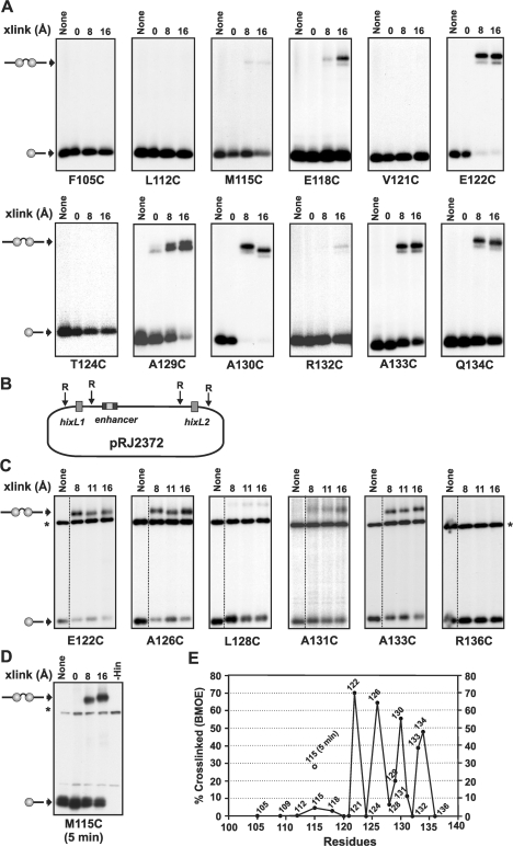
Figure 6.
Crosslinking of helix E cysteine mutants. (A) Crosslinking reactions were performed using 3′ 32P-labeled 36 bp hixL fragments. Crosslinked synaptic complexes were purified from native polyacrylamide gels, extracted and then subjected to SDS–PAGE. The A129C panel is from referece (20). (B) Diagram of pRJ2372. (C) Crosslinking reactions performed using supercoiled pRJ2372 in the presence of Fis. After 1 min crosslinking reactions, the plasmid was digested with EcoR1, which cleaves 50 bp on either side of each hix site, labeled with 32P-ATP, and subjected to SDS–PAGE. The labeled bands denoted with an asterisk and present in all lanes are from a DNA fragment released by EcoR1 cleavage. All crosslinking reactions in A and B were performed for 1 min using diamide (0 Å), BMOE (8 Å spacer), BMB (11 Å) or BMH (16 Å spacer). The Hin–M115C reaction in (D) was crosslinked for 5 min. Each cysteine was introduced into a Hin–H107Y background. (E) Plot of crosslinking efficiencies of cysteines as a function of location along helix E. Percentage crosslinked is the amount of crosslinked di-protomer/non-crosslinked + crosslinked Hin–DNA covalent complexes. Data is from 1 min BMOE crosslinking reactions on supercoiled DNA. The results of 5 min BMOE crosslinking reactions with M115C (D) is also denoted.

