Figure 3.
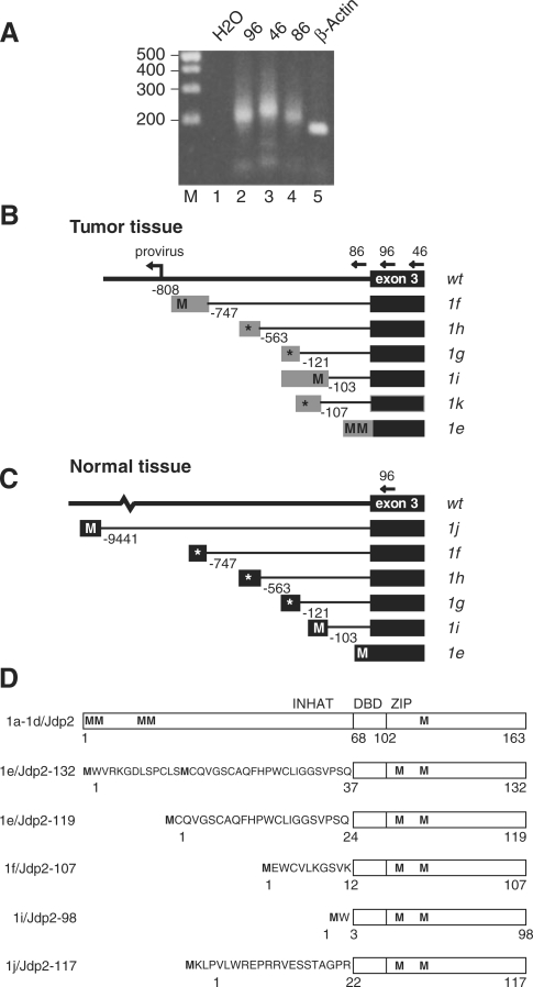
Identification of Jdp2 intron 2 mRNAs. (A) Ethidium bromide-stained agarose gel showing representative PCR products with linker-specific forward primer on 5′ RACE cDNA from tumor 1161 using different reverse primers in Jdp2 (oligos 96, 46 and 86, lanes 2, 3 and 4, respectively) and Actb exon 3 (lane 5); in lane 1 no cDNA template was added. (B) and (C) Schematic structure of the alternative Jdp2 exon 1e through 1k as found by 5′ RACE in tumor tissue (B) and normal tissue (C). Positions of exon 1-specific splice donor sites relative to exon 3 are given in base pairs. Putative start codons (M) in frame with the ORF of Jdp2 are indicated, while an asterisk indicates that no ORF is present in frame with Jdp2. (D) Protein structure of Jdp2 as generated from exon 1a through 1d, and predicted Jdp2 isoforms generated from exon 1e, 1f, 1i and 1j. The INHAT domain as well as the basic DNA binding domain (DBD) and the leucine zipper (ZIP) regions are indicated. Methionines are indicated (M) and the N-terminal peptides are shown for the isoforms.

