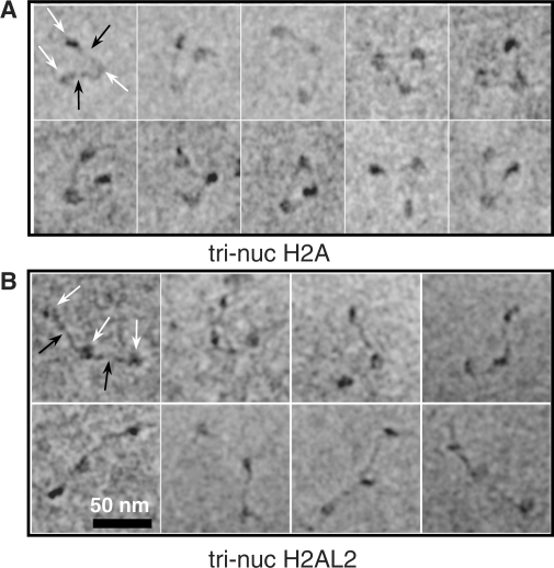
Figure 6.
Electron cryo-microscopy visualization of conventional H2A and histone variant H2AL2 tri-nucleosomes. A DNA fragment containing three tandem 601 positioning sequence repeats was used to reconstitute conventional (A) and H2AL2 tri-nucleosomes (B) and they were visualized by E-CM. Typical micrographs for both types of particles are shown. The conventional trinucleosomes exhibit V-shaped structure with the two-end nucleosomes at both ends of the ‘V’ and the middle nucleosome at the center of the ‘V’. In contrast, the majority of the H2AL2 trinucleosomes exhibit ‘beads on a string’ structure and very few H2AL2 trinucleosomes show open ‘V’-type of organization. Black arrows indicate the linker DNA, while the nucleosome is designated by white arrows.

