Figure 2.
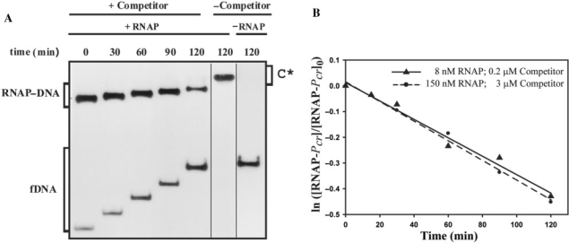
Kinetic of dissociation of RNAP from Pcr at 37°C. (A) EMSA analysis of the stability of the RNAP–Pcr complexes. Equilibrium mixtures contained 150 nM RNAP and 2 nM labelled DNA. Samples were analyzed at the indicated times following addition of 3 μM unlabelled DNA. Bands corresponding to free DNA (fDNA) and to specific RNAP–DNA complexes are indicated. On addition of competitor (t = 0), the complex fraction was estimated to be 0.8. In the absence of competitor, slower-migrating complexes (C*) that contained several RNAP molecules were seen. All the lanes displayed came from the same gel. (B) Time course of RNAP–Pcr complex dissociation. Linear fits of data from each of two experimental conditions are shown.

