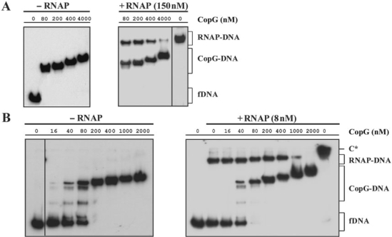
Figure 5.
CopG-concentration-dependent dissociation of the RNAP–Pcr complexes at 37°C. RNAP (at the concentrations indicated in A and in B) and DNA (2 nM) were equilibrated at 37°C and then treated with heparin (150 μg/ml in A, and 10 μg/ml in B) before adding different amounts of CopG. Incubation of the mixtures continued for 10 more minutes before loading onto the gel. CopG–DNA complexes formed in the absence of RNAP were also analyzed. Bands corresponding to free DNA (fDNA) and to RNAP–DNA and CopG–DNA complexes are indicated. At high CopG concentrations (>200 nM), complexes migrating slower than the specific complex generated by binding of four CopG dimers to the operator DNA (17) were observed. These slowly-migrating complexes contain additional repressor molecules nucleated from the operator (39). All the lanes displayed came either from the same gel or from gels prepared and run in parallel. Only one of two independent experiments yielding identical results is shown in each panel. Note that in the absence of heparin, slowly migrating RNAP–DNA complexes that contained several RNAP molecules were seen (C* in panel B).

