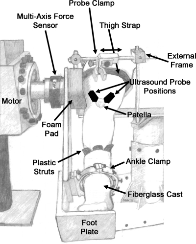
Fig. 1.
Experimental setup. The subject was seated in a custom chair with the hip at 85° and the knee at 60°. The plantar surface of the foot was supported on a foot plate. Rigid plastic struts were placed on the medial, lateral, and anterior aspects of the ankle joint inside a fiberglass cast. The fiberglass cast covered the midfoot and ankle and was secured to the footplate using a custom clamp. The clamp was tightened to ensure no translation or rotation of the tibia during isometric knee extension. The ankle clamp and foot plate were attached to a six degree-of-freedom torque sensor through an aluminum beam. An external frame was used to fix the ultrasound probe over the tendon of interest. The black ovals on the knee represent the approximate position of the ultrasound probe for measurement of vastus medialis obliquus (VMO) and vastus lateralis (VL) tendon elongation, slightly overlapping the proximal patella.

