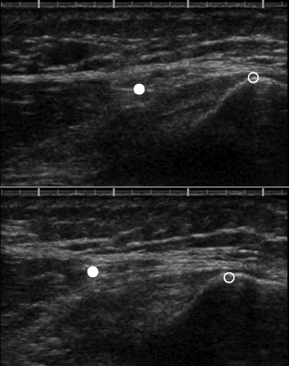
Fig. 2.
Measurement of VMO tendon elongation. The ultrasound probe was positioned such that the angle of orientation was maintained and the proximal portion of the patella (open circle) was visible in the image. For the VMO tendon, the myotendinous junction (MTJ; filled circle) was also visible in the image. Top: ultrasound image before isometric quadriceps contraction. Bottom: image during isometric quadriceps contraction (7 N·m). Notice the proximal shift of both the patella and the MTJ during contraction; larger proximal shift of MTJ indicates tendon elongation. Proximal shift of the patella is due to patellar tendon elongation.

