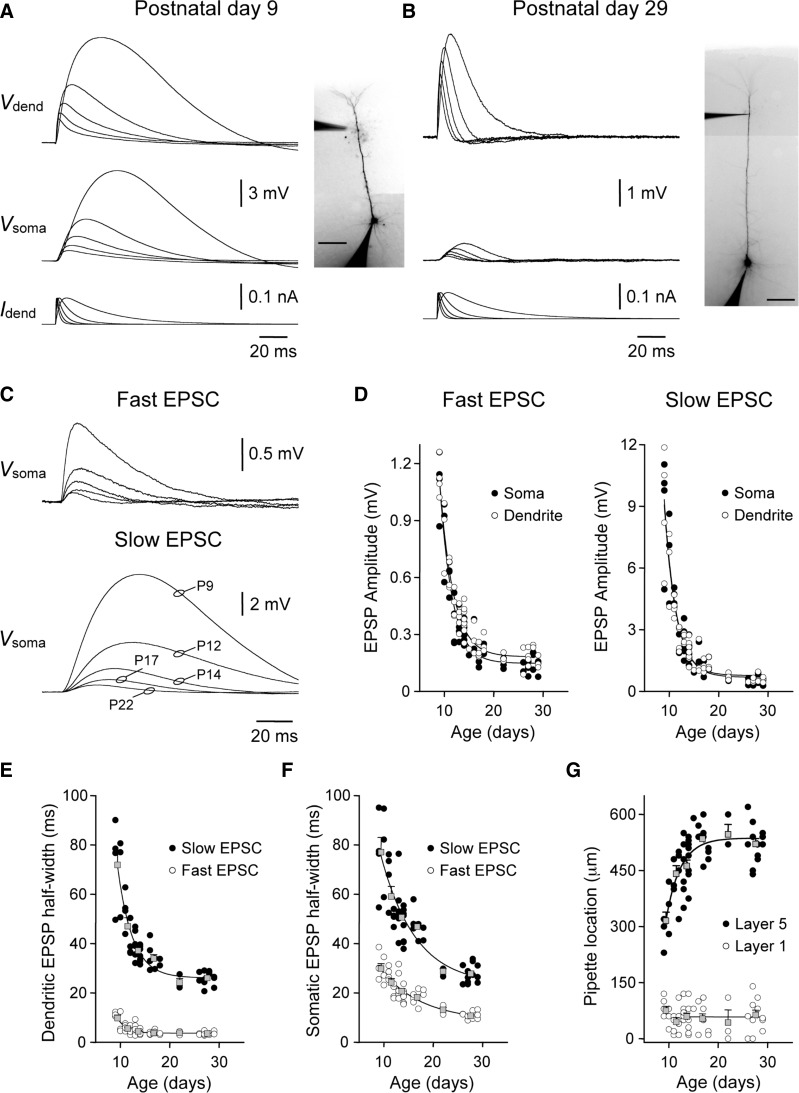
FIG. 1.
Developmental decrease in the somatic impact of apical dendritic excitatory postsynaptic potentials (EPSPs). A and B: simultaneous somatic (Vsoma) and dendritic (Vdend) recording of simulated EPSPs in a postnatal day 9 (P9, A) and P29 (B) layer 5 pyramidal neurons. Simulated EPSPs were generated by the injection of a series of simulated excitatory postsynaptic currents (EPSCs) at the distal apical dendritic recording site (Idend; bottom traces). The amplitude and kinetics of simulated EPSPs are transformed with development. Neuronal morphology and the placement of recording electrodes are shown in the inset photomicrographs (scale bars = 100 μm). C: age-dependent reduction in the somatic amplitude of distal dendritically generated EPSPs. Overlain traces show simulated EPSPs recorded at the indicated postnatal ages. Fast and slow EPSCs had τrise of 0.2 and 3 ms and τdecay of 2 and 30 ms, respectively. D: pooled data show the age-dependent decrease in the amplitude of EPSPs, generated with fast and slow EPSC kinetics, when generated at the soma and recorded at an apical dendritic site (○) or vice versa (•). Data have been fit with single exponential functions (—). E and F: developmental speeding of the kinetics of simulated EPSPs generated by fast or slow EPSCs. Simulated EPSPs were recorded at their site of generation (E) and following spread to the soma (F). Overlain gray symbols show results pooled into 6 age groups (means ± SE). Grouped data have been fit with single-exponential functions (—). G: position of recording electrodes. •, the distance of the apical dendritic recording electrode from the soma of layer 5 pyramidal neurons; ○, the distance of the dendritic recording electrode from the layer 1–layer 2 border. Grouped data have been fit with a single-exponential function (layer 5) or linear regression (layer 1).

