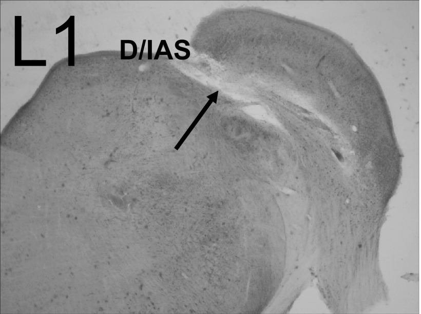
FIG. 5.
Photomicrograph of a lesion in the D/IAS (L1). An injection of 0.2 μl melittin into the region of the D/IAS, produced a lesion with a rostral-caudal extent of 560 μm that damaged only the D/IAS (arrow). Electrophysiological results from this animal are depicted in Fig. 6, A and B.

