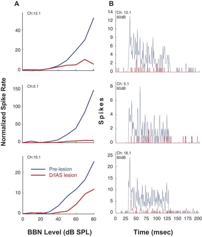
FIG. 6.
A: rate-level functions for 3 sorted single units from 1 animal to contralateral sound after cochlear ablation. Responses are shown before and after a melittin lesion in the D/IAS (L1). Spike rate is normalized to spontaneous rate. B: PSTHs of single-unit responses to contralateral sound after cochlear ablation (bin width, 1 ms). Responses shown are pre- and post-lesion (as indicated). Melittin lesion is in the D/IAS (L1). Photomicrographs of the lesion in this animal are shown in Fig. 5. Stimulus: 100-ms duration BBN.

