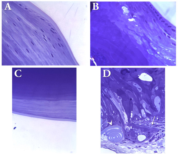
Figure 6.
Posterior lens defects in R49Cneo knock-in mouse. Images are shown for lens sections from 9.5-month-old WT/WTneo and WT/R49Cneo mouse. (A) Brightfield image of the equatorial region in a WT/WTneo mid-sagittal lens section. (B) Brightfield image of the equatorial region in a WT/R49Cneo mid-sagittal lens section. (C) Posterior region of a WT/WTneo lens shows a normal posterior capsule. (D) A WT/R49Cneolens with posterior cataract. Note the extensive posterior rupture, migration of cells and cell debris (arrow), and compaction of the ruptured posterior capsule (arrowhead).

