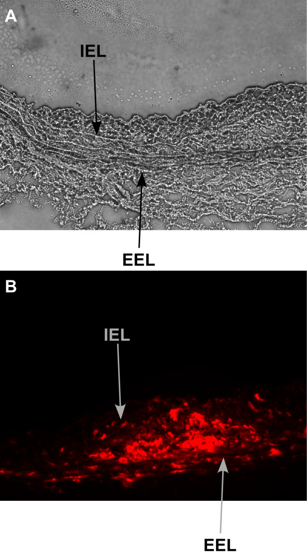
Figure 2.
Tracking of delivered cells. Light microscopy cross section (20×) showing neointima formation in immunodeficient rat carotid 4 weeks after balloon injury (A). CM-Dil-labeled human CD34+ cells stain red under fluorescent microscope (20×) within intima and media of carotid 4 weeks after balloon injury (B). IEL = Internal elastic lamina, EEL = external elastic lamina.

