
Figure 1.
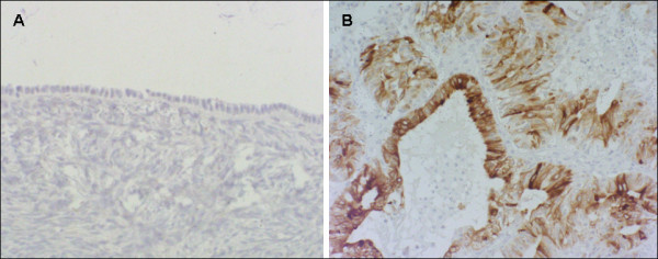
Representive immunohistochemical staininig for MGB-2 in normal ovary and ovarian adenocarcinoma. Celomic epithelium and parenchyma of normal ovary are negative for MGB-2 (A). Early-staged ovarian adenocarcinoma with endometrioid histology showing a staining intensity variable from moderate to strong (B).