
Fig. 8.
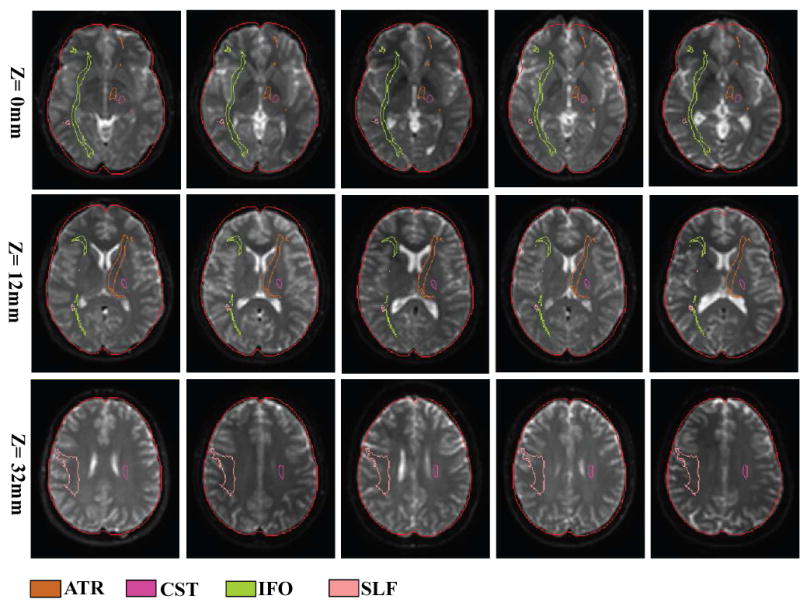
Normalized images of subject #1, 3, 5, 7, 9 used in Fig. 4 – 6 to demonstrate registration quality. Three axial slices at z = 0, 12, and 32 are shown, which reveal the probabilistic locations of the IFO (green), the SLF (peach), the ATR (orange), and the CST (pink). The outer boundary defines the shape of the ICBM-152 template.