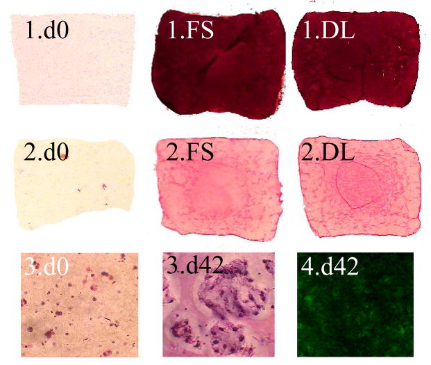
Figure 6.
(1) Safranin O staining for GAG, (2) Picrosirius Red staining for collagen, (3) hematoxylin and eosin staining for visualization of local multiplication of cell nuclei (Mag. 40x), and (4) Immunohistochemical staining for type II collagen. All slides taken from study 3 on either day 0 or day 42 with either free-swelling (FS) or dynamically-loaded (DL) groups.

