
Figure 4. Confirmation of siRNA knockdown of FKBP52 and FKBP51 in SH SY5Y cells.
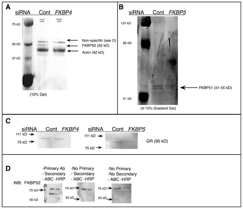
Differentiated SH-SY5Y cells on glass coverslips in 24 well plates were exposed to 250 ng siRNA generated against the FKBP4 (a) gene or against FKBP5 gene (b) and a control gene, encoding GFP provided by the manufacturer, for 24 hr; protein was isolated for Western blot. Concurrent to probing for FKBP52 a western blot for Actin was performed for the loading control (a). To ensure that a difference in GR immunofluorescence was not due to altered GR protein content, GR was assessed by western analysis (c). To discern non-specific bands in the western blots, membranes were developed with primary antibody (d-left), without primary antibody (d-middle) and without either secondary or primary antibody (d-right center), and it was determined that the non-specific band originates from the avidin-biotin amplification system in (a).