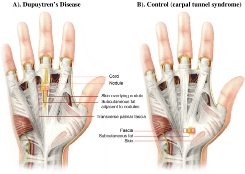
Figure 2.
Tissues subjected to analysis in this study. a This figure demonstrates the palm of the hand of an individual affected with Dupuytren’s disease, where the overlying skin has been removed to demonstrate the position of the palmar fascia in relation to the disease and harvested samples as indicated. Skin overlying palmar nodule, subcutaneous fat adjacent to palmar nodule, and transverse palmar fascia (Skoog’s fibers) were obtained from Dupuytren’s disease patients. b This figure demonstrates the palm of the hand of a control subject, where the overlying skin has been removed to demonstrate the position of the palmar fascia harvested. Skin, palmar fascia (transverse carpal ligament), and fat were obtained from control subjects, individuals undergoing carpal tunnel release.

