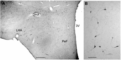
Fig. (2).
Distribution of Hcrt/Orx neurons in the cat hypothalamus. A: Microphotograph of a coronal section of cat hypothalamus showing the distribution of orexinergic neurons as result of the immunoreaction for anti-Orexin A antiserum. No counterstaining. B: High magnification of area squared in A. DHA. dorsal hypothalamic area, LHA: lateral hypothalamic area, PeF: perifornical region, 3V: third ventricle. Calibration bars: A, 500 µm, B, 100 µm.

