
Fig. 6.
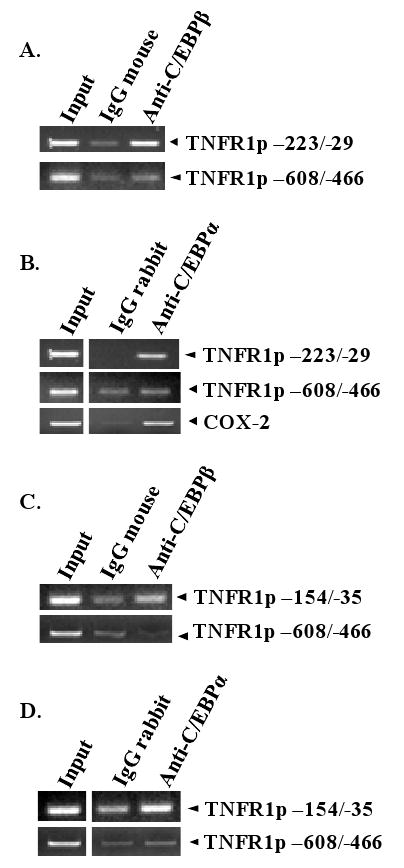
C/EBPα and C/EBPβ bind to the TNFR1 promoter in vivo. ChIP assays were performed in HONE-1 (A, B) and AGS-B95.8 (C, D) cells using control IgG mouse antibody, mouse anti-C/EBPβ antibody, control rabbit IgG antibody, or rabbit anti-C/EBPα antibody. PCR was performed using two sets of primers for the TNFR1 promoter encompassing the region containing the C/EBP binding motif (-223/-29 and -154/-35), a region of the TNFR1 promoter which does not bind to C/EBP (-608/-466), or a region of the COX-2 promoter known to bind to C/EBPα. Input DNA for each condition is shown in the first lane.