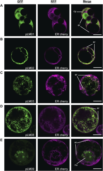
Fig. 6.
Co-localization experiments in Arabidopsis root protoplasts. Protoplasts were co-transformed using the constructs presented in Fig. 1 and an ER marker protein fused to RFP, ER cherry (Nelson et al., 2007). (A) pLM01 (GFP control). (B) pLM02 (BSP-GFP). (C) pLM03, (BSP-LT-B::GFP). (D) pLM08 (ZSP-LT-B::GFP). (E) pLM09 (LT-B::GFP). Green channel corresponds to GFP signal. Red channels (presented in magenta color) corresponds to ER-cherry signal. Merged images are also presented. Organelle labelling: cytosol (cy), nucleus (nu), endoplasmic reticulum (er). Bars=10 μm.

