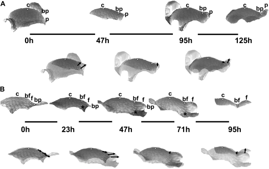
Fig. 5.
Sequences of 3D reconstructions of replicas taken from two individual shoot apices of Anagallis arvensis in the reproductive developmental phase, representing consecutive stages of bract and flower primordium development: initial bulging, lateral expansion, and separation stages of bract primordium development (A); lateral bulging, separation, and axil deepening stages of flower primordium development (B). White dots on the profiles point to: c, the cell on the top of the SAM; bp, the cell at the boundary between the meristem and bract primordium; p, the cell on the bract primordium tip; bf, the cell at the boundary between the flower primordium and the SAM; and f, the cell at the tip of the flower primordium. Asterisks point to the cell at the base of the bract subtending the flower primordium. Other labelling is as in Fig. 4.

